Directing Clinical Research in Academic-Industry Partnerships
Ming is the Visage Imaging, Inc. Director of Clinical Research for North America and is stationed at the Yale-New Haven Hospital (Associate Professor Adjunct, Radiology and Biomedical Imaging) where he oversees, coordinates, drives and directs research collaborations with high profile academic hospitals in North America to develop new solutions for diagnostic image analysis and guidance that improve clinical and operational outcomes while reducing cost of care. This includes applications of Artificial Intelligence (AI) and Machine Learning (ML) in the Radiology enterprise diagnostic imaging solutions space. Ming identifies opportunities for academic-industry research partnerships, and acts as the liaison between Visage Imaging researchers and clinical collaborators to translate ideas to prototype for clinical validation, with the goal of technology transfer to product. A highlight is Ming coordinated the clinical data curation and ground-truth annotation for building a fully automatic breast density AI classifier that provides an ACR BI-RADS Atlas 5th Edition breast density category to aid radiologists in the assessment of breast tissue composition from full field digital mammography and digital breast tomosynthesis systems and drove the clinical validation with Yale radiologists and Visage developers that led to transfer to product and regulatory approvals in 21 months following IRB approval and has been in full clinical production use at Yale since April 2021. Moreover, Dr. Lin directed the study with Yale radiologists to assess the AI algorithm’s post-clinical deployment performance, and we found there was 99.35% agreement in classifying the breast density between the AI and the radiologist. This was the first FDA-cleared AI algorithm that reported having >1000 patients for validating the AI from two different clinical sites: Yale and New York University (NYU), and it was the first for a major PACS vendor to offer a self-developed, FDA-cleared AI algorithm natively into their PACS: Visage Breast Density, K201411, 510(k) clearance, January 2021, Health Canada Licensed, October 2020, Australian TGA approval, July 2020, CE Mark Cleared, May 2020. Another effort Dr. Lin is working on is multi-institution AI research to develop robust deep learning methods for generating patient-specific virtual-high-count PET images from standard PET images, thereby saving imaging time, reducing radiation dose, and increasing scanner longevity. This work is being conducted in an NIH R01 academic-industry partnership grant where Dr. Lin is the Visage Imaging, Inc. Principal Investigator (PI), and the other partners are Yale New Haven, Massachusetts General Brigham, and University of California Davis hospitals.
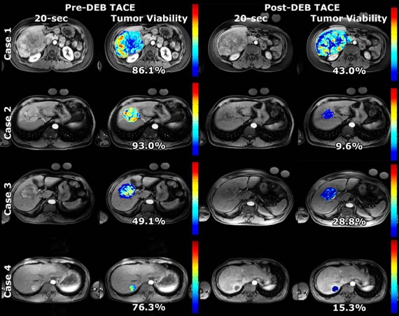
Dr. Lin also is directly involved in research to develop better ways to treat patients with liver cancer using transcatheter arterial chemoembolization (TACE) and in this context, Ming is also the chief engineer and operations manager of the Yale Interventional Oncology Research Lab. Dr. Lin is Principal Investigatoron two NIH R01 grants and the Industry PI on its renewal NIH R01 grant (three grants in total) to improve the diagnosis, treatment, and response assessment after transcatheter arterial chemoembolization for patients with liver cancer. A highlight is the development of a 3D quantification method for improved response assessment after TACE called quantitative European Association for the Study of the Liver (qEASL). Ming's invention has been shown to be much improved over the current tumor viability measurement methods that are qualitative and 1- or 2-dimensional in being a more accurate match to pathology and in predicting patient survival sooner so that treatment decisions can be made earlier. Ming's prototype was validated and shown to have clinical value and transferred to be come a product that received FDA 510(k) approval.
Prior to Visage Imaging, Ming was the Philips Healthcare research site manager and senior researcher stationed onsite at Yale where he managed the research portfolio and partnership Philips has with Yale. Ming obtained his Ph.D. in biomedical engineering at Duke University researching high resolution x-ray imaging for small animals. The research was conducted at the Duke Center for In-Vivo Microscopy.